What you’ll learn:
- Why 850-nm light is well-suited to deep-body hemoglobin sensing.
- How VCSELs are used to create ultrasonic signal returns.
- How the combined optical/acoustic array was arranged and fabricated.
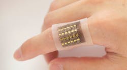
By combining two very different types of transducers in a soft, skin-friendly substrate, a team of engineers at the University of California (San Diego) devised an electronic patch that can monitor biomolecules such as hemoglobin in deep tissues. As a flexible, low-form-factor wearable patch, it’s easily and comfortably attached to the skin, allowing for monitoring that’s long term and noninvasive—a winning combination.
It can perform three-dimensional mapping of hemoglobin with a submillimeter spatial resolution even centimeters below the skin, in sharp contrast to other wearable electrochemical measurement units that can only sense critical biomolecules on the skin surface or immediately below it. This is useful as it should be able to detect, among other concerns, low blood perfusion inside the body (perfusion is the local fluid flow through the capillary network and extracellular spaces, and is necessary for the transport of oxygen, nutrients, and waste products).
This malady may cause severe organ dysfunctions. It’s associated with a range of ailments including heart attacks and vascular diseases of the extremities, while abnormal blood accumulation in areas such as in the brain, abdomen, or cysts can indicate cerebral or visceral hemorrhage or malignant tumors. As a result, this patch could help spot life-threatening conditions such as malignant tumors, organ dysfunction, cerebral or gut hemorrhages, and more.
The patch is equipped with a dual array of laser diodes and piezoelectric transducers in its soft silicone polymer matrix. The vertical-cavity surface-emitting laser (VCSEL) diodes direct pulsed optical beams into the tissues, and biomolecules in the tissue absorb the optical energy.
Unlike conventional ultrasound systems where the piezoelectric transducers create and then receive the ultrasound waves, here these transducers act as “receive only” sensors. The optical signals, which can penetrate over 2 cm into biological tissues, activate hemoglobin molecules to generate acoustic waves that are collected by the transducers for 3D imaging of the hemoglobin with a high spatial resolution.
Patch Design and Fabrication
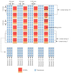
In the patch, 24 VCSELs are evenly distributed in four equally spaced columns to generate uniform illumination in regions below the patch. The 240 individually addressable piezoelectric transducers are arranged in between the VCSELs, in 15 columns with 16 transducers in each column (Fig. 1). The team even analyzed how VCSEL distribution affects the imaging performance.
In a project such as this, transducer specifics are critical. High-power 850-nm VCSELs are used to achieve the needed detection depth and a high signal-to-noise ratio. This wavelength has multiple virtues:
- It performs deep-tissue penetration.
- It’s in the first optical window for probing human tissue.
- Hemoglobin also has the dominant optical absorption coefficient at this wavelength compared with other molecules, such as water and lipid.
- VCSELs at 850-nm wavelength are very common and widely used with optical fibers.
- The complementary photodetectors are widely available and low cost.
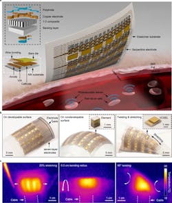
The receiving transducer element is composed of a piezoelectric layer and a backing layer made of 2-MHz lead-zirconate-titanate (PZT) micropillars embedded in epoxy (Fig. 2). In contrast to bulk PZT, this composite suppresses the transverse vibration and enhances the axial vibration of the PZT micropillars. This increases the electromechanical coupling coefficient and improves the energy-conversion efficiency.
The resultant soft photoacoustic patch is mechanically and electrically robust, even under different modes of deformation including bending, wrapping, twisting, and stretching. While the patch is rigid locally at each piezoelectric transducer element and laser diode, it’s soft globally at the system level, and no external pressure is required to conformally attach it to the skin. Further, mechanical deformations don’t affect the performance of the VCSELs.
Testing Results
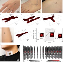
Among the many tests, they evaluated the performance specifics in vivo on various body parts of a live subject (the inherently non-risky, non-invasive nature of the patch is a major advantage here) (Fig. 3). The results were in close agreement with the anticipated performance as well as simulations.
They also plan to explore the wearable patch’s potential for core-temperature monitoring, as the photoacoustic signal amplitude is proportional to the temperature. They have demonstrated the ability to do so via some preliminary ex-vivo experiments.
The work was supported in part by grants from the Air Force Research Laboratory and National Institutes of Health. It’s presented in a detailed, 13-page, highly readable paper “A photoacoustic patch for three-dimensional imaging of hemoglobin and core temperature” (with 24 authors!) published in Nature Communications. If that’s not enough, there’s considerably more detail covering all aspects in their posted 84-page associated Supplementary Information file.


